Arterial
The arterial system constitutes the high-pressure efferent conduit tree branching from the left ventricle (aorta) and right ventricle (pulmonary trunk) delivering pulsed blood into microvascular capillary beds.
Quantitative Metrics & Physiological Scale
Wide high-volume outflow conduit directly accepting stroke volume
Physiological hydraulic pressure generated during left ventricular ejection
Tunica intima, elastica media, and adventitial connective tissue
Aortic elastic recoil sustaining diastolic blood perfusion pressure
Physiological Role & Homeostasis
High-pressure distribution of oxygen-saturated blood from left ventricle to peripheral organs
Peripheral vascular resistance regulation through arteriolar smooth muscle contraction
Systolic pulse pressure buffering transforming intermittent stroke boluses into continuous flow
Elastic Arteries vs Muscular Arteries
| Feature / 比较维度 | Elastic Arteries / 弹性动脉 (主动脉/颈总/锁骨下) | Muscular Arteries / 肌性动脉 (桡/股/脾/肠系膜动脉) |
|---|---|---|
| Tunica Media Composition / 中膜主要组织学构成 | Multiple dense concentric elastic laminae / 多达数十层厚密同心弹性板膜,平滑肌含量相对较少 | Abundant circular smooth muscle cells (up to 40 layers) / 丰厚的环行平滑肌层,弹性纤维相对退化 |
| Functional Specialization / 核心生理分工 | Pressure damping and pulse wave conduction (Windkessel) / 缓冲收缩压震荡、维持舒张压平稳搏动 | Active vasomotion and targeted organ flow distribution / 主动血管舒缩调控,根据器官代谢率调整灌注阻力 |
| Lumen Caliber & Pressure Wave / 管腔口径与脉搏波特征 | Large lumen (>10 mm); low hydraulic flow resistance / 口径宽大(>10 mm);极低水力流阻,高顺应性 | Medium caliber (2-10 mm); responsive to sympathetic tone / 中等口径(2-10 mm);对交感神经去甲肾上腺素极度敏感 |
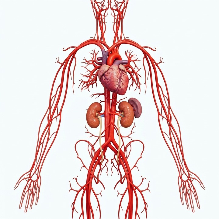
Full 3D Atlas Coverage
This system is fully interactive in the 3D Full-Body Atlas with complete anatomical parts, layer visibility toggling, and exploded view.
How to Study This System in 3D
Trace the three classic aortic arch branches: brachiocephalic trunk, left common carotid, and left subclavian artery.
Observe how major abdominal branches (celiac trunk, superior/inferior mesenteric) project to visceral organs.
Explore Synergistic Anatomical Systems
For educational use only; not medical advice, diagnosis, or treatment.